For decades, radiologists have relied on two-dimensional slices to interpret complex anatomy. That was a skill demanding immense precision and experience. Yet, in many cases, subtle lesions, overlapping structures, or ambiguous planes make diagnosis a challenge, especially when clinical urgency demands faster reporting. This is where 3D imaging has transformed radiology’s landscape.
By turning flat data into lifelike visualizations, 3D imaging allows clinicians to explore anatomical details from every angle, offering depth, context, and clarity that traditional scans often lack. When combined with Multi-Planar Reconstruction (MPR) tools, it bridges the gap between visualization and understanding, revealing nuances that guide accurate diagnoses and better patient outcomes.
In this blog, advanced radiology software and teleradiology systems are empowering radiologists to view, analyze, and report with greater confidence, marking a new era in diagnostic precision.
What is Multi-Planar Reconstruction (MPR)?
Multi-Planar Reconstruction (MPR) is one of the most transformative developments in modern radiology software that enables radiologists to visualize medical scans beyond the traditional axial view. It allows 2D cross-sectional images, typically from CT or MRI, to be reformatted into additional planes such as sagittal, coronal, or even oblique orientations. This layered reconstruction gives clinicians the ability to explore internal structures with far greater clarity, improving understanding of anatomical relationships, and subtle pathological changes.
In clinical settings, MPR plays a crucial role in cases where precision and spatial awareness are vital, from evaluating vascular structures to assessing complex fractures or tumor boundaries. By generating dynamic, multi-view reconstructions, it ensures no diagnostic information is lost between slices. For radiologists, MPR becomes the bridge between raw imaging data and a comprehensive diagnostic perspective, enhancing both efficiency and accuracy.
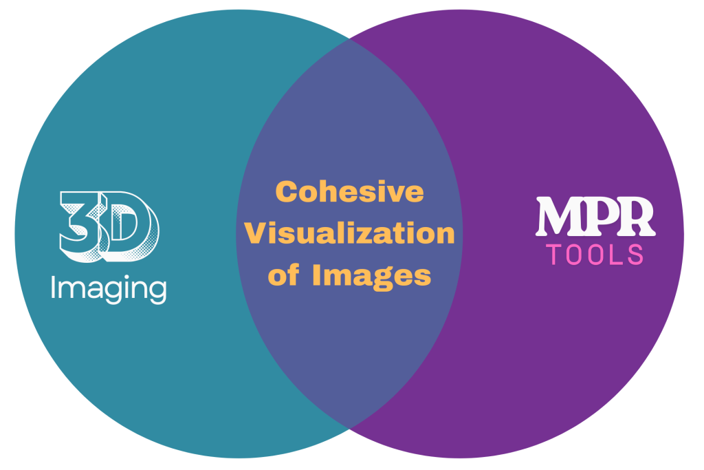
Then, How 3D Imaging & MPR Work Together?
When combined, 3D imaging and MPR tools transform raw scan data into a cohesive, spatially accurate visualization, offering radiologists a complete, interactive view of the human body.
- Integrated Visualization: MPR provides foundational planes, while 3D imaging compiles these slices into a volumetric model for intuitive viewing.
- Depth and Detail: Radiologists can rotate, zoom, and navigate through 3D-rendered anatomy, ensuring nothing remains hidden within flat cross-sections.
- Precision Diagnosis: The combination helps detect minute lesions, vessel anomalies, or soft-tissue variations that are easily missed in 2D scans.
- Workflow Harmony: When embedded into PACS or teleradiology software, the fusion of MPR and 3D imaging accelerates reporting without compromising accuracy.
- Clinical Collaboration: 3D-MPR outputs enhance communication between radiologists, surgeons, and clinicians, turning image data into actionable clinical insight.
How 3D Visualization Enhances Diagnosis
Modern radiology is no longer limited to viewing stacked slices on a screen. This deeper, interactive visualization is not just a technical advancement; it’s a clinical advantage that directly impacts diagnostic confidence and patient outcomes.
Below are some ways 3D visualization elevates diagnostic workflows and decision-making across specialties:
1. Enhanced Anatomical Clarity
3D imaging converts routine CT or MRI data into interactive models, allowing radiologists to examine tissues, organs, and pathologies from multiple perspectives. It helps differentiate overlapping structures, for example, distinguishing a vascular loop from a mass in neuroimaging or clarifying bone fragment alignment in trauma cases. This precision leads to more consistent interpretation across departments.
2. Accurate Lesion Localization & Measurement
In traditional imaging, locating a lesion’s exact position can be time-consuming. With volumetric 3D medical imaging, radiologists can pinpoint its depth, measure dimensions accurately, and understand spatial relationships with surrounding tissues, essential for pre-surgical planning and oncology staging.
3. Streamlined Collaboration Between Teams
3D visualization serves as a universal language across medical disciplines. Surgeons, oncologists, and referring physicians can review the same 3D-rendered model, gaining a clearer understanding of the pathology. This shared visual context improves communication, shortens reporting cycles, and minimizes discrepancies between clinical and imaging interpretations.
4. Faster and More Confident Decision-Making
With interactive radiology visualization tools, radiologists can rapidly navigate through datasets, assess key findings, and finalize reports more efficiently. The ability to correlate 3D reconstructions with Multi-Planar Reconstruction (MPR) views also ensures that critical findings aren’t overlooked, reducing the need for repeated scans or second opinions.
5. Improved Follow-up and Case Comparison
When tracking disease progression or post-surgical recovery, 3D imaging tools provide measurable data over time. Volume-based comparisons reveal subtle changes that might be missed on 2D slices, helping clinicians monitor therapeutic response with confidence and precision.
In essence, 3D visualization represents a turning point for advanced diagnostic imaging, bringing together clarity, speed, and collaboration into a unified diagnostic workflow that empowers both radiologists and clinicians to deliver better patient care.
Integration in Modern Radiology Systems
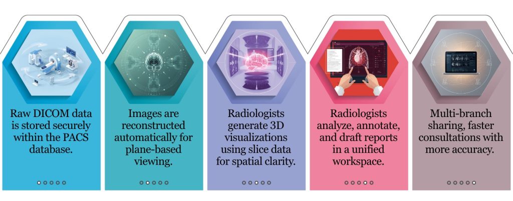
The real strength of 3D imaging lies not only in its visual capabilities but in how seamlessly it fits within the modern radiology workflow. When integrated into PACS and teleradiology systems, 3D and Multi-Planar Reconstruction (MPR) tools transform the way clinicians review, interpret, and share imaging data. Instead of switching between applications or formats, radiologists can now reconstruct, analyze, and report all within a single, connected environment..
This integration ensures faster data retrieval, consistent visualization quality across branches, and greater efficiency in both reporting and collaboration. It also allows hospitals and diagnostic centers to maintain centralized access to imaging data, making multi-department consultations, second opinions, and remote interpretations simpler and more accurate.
Mediog: Powering the Next Generation of 3D Diagnosis
Mediog’s focus has always been on simplifying radiology without compromising clinical depth. Our cloud-based platform integrates 3D imaging and MPR tools directly into the diagnostic workflow, that allows radiologists to move effortlessly from image acquisition to interpretation and reporting. Every 3D visualization is generated with precision, ensuring consistency across studies and branches, whether viewed from a hospital workstation or a remote reporting setup.
The best thing is that, Mediog’s teleradiology software supports multi-modality data, structured reporting, and AI-assisted rendering. Now radiologists will have full control over visualization of quality and workflow efficiency. The system is designed to adapt to hospital-specific protocols, helping clinicians maintain accuracy and compliance while achieving faster turnaround times.
Trusted by leading healthcare institutions in Kolkata like Desun Hospital, Apollo Diagnostics, and Suraksha Clinic & Diagnostics, Mediog continues to strengthen the bridge between advanced technology and patient-focused diagnosis, quietly transforming how radiology works, every single day.
Conclusion
3D imaging and MPR have reshaped radiology, bringing clarity, speed, and confidence to every diagnostic step. As imaging grows more data-driven, the ability to visualize in three dimensions isn’t just an advantage; it’s essential for precise, patient-centered care. With Mediog’s integrated 3D-ready platform, radiologists can experience this advancement firsthand, optimizing workflows and ensuring every diagnosis reflects the highest standard of clinical accuracy.
###